BrainLab Airo Mobile Intraoperative CT Scanner System
Technology
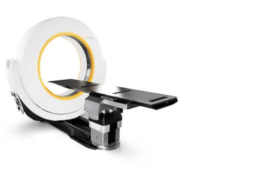
The BrainLab Airo Mobile Intraoperative CT scanner system is a mobile imaging platform that can be used during surgery to obtain a high-resolution CT scan of a patient while in the operating room undergoing surgery.
Specifically, the technology uses ultrasound waves to create a small ablation to disrupt pathological brain activity in order to reduce the signs or symptoms of Essential Tremor and Tremor-dominant Parkinson’s Disease.
Who can benefit
Patients undergoing electrode implantation into the brain, such as for epilepsy or for deep brain stimulation.
Patients who are having spinal instrumentation placed, or a spinal tumor resected, may benefit from an intraoperative CT scan. By doing a CT scan in the operating room, your surgeon can ensure that your spinal instrumentation was accurately placed, and perform any revisions needed before you leave the operating room.
BrainLab AIRO mobile CT scanner with navigation system can create an up-to-date 3D model of your spine. Your surgeon can use that model to help accurately place spinal instrumentation, or ensure maximal tumor resection.
How it works
BrainLab Airo Mobile Intraoperative CT scanner is brought into the surgical room prior to your surgery. The operating room table, where your surgery is performed, is attached to the CT scanner. A CT scan is performed prior to utilizing the Brainlab navigation system to register your brain / spine to a reference array placed on a fixed location on your body, typically your spine or hip. If you have spine hardware placed or a tumor resected, a CT scan is performed after the surgery is complete, but just prior to closure of your wound. This is to verify that any spine hardware placed is in the correct location, and if a tumor is resected, whether there is any residual left. Once the surgeon is satisfied with the CT result, your wound is closed.
 The BrainLab Airo Mobile Intraoperative CT scanner system is a mobile imaging platform that can be used during surgery to obtain a high-resolution CT scan of a patient while in the operating room undergoing surgery.